File:Fluorescence in situ hybridization of plasmablastic lymphoma.jpg


No higher resolution available.
Fluorescence_in_situ_hybridization_of_plasmablastic_lymphoma.jpg (320 × 250 pixels, file size: 9 KB, MIME type: image/jpeg)
(Fluorescence in situ hybridization study performed on interphase cells using probes to the MYC (red signal) and IGH (green signal) loci and chromosome 8 centromere (blue signal) demonstrates that the neoplastic cells contain a reciprocal translocation between the MYC and IGH loci (yellow signals, arrows); image and legend from Boguscz et al. Am J Clin Pathol. 2009.)
File history
Click on a date/time to view the file as it appeared at that time.
| Date/Time | Thumbnail | Dimensions | User | Comment | |
|---|---|---|---|---|---|
| current | 12:35, 20 May 2020 | | 320 × 250 (9 KB) | Mark.Evans (talk | contribs) | (Fluorescence in situ hybridization study performed on interphase cells demonstrating rearrangement of MYC (yellow signals); image courtesy of Fabiola Quintero-Rivera, MD) |
| 10:40, 30 April 2020 | 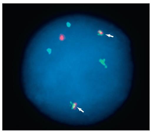 | 520 × 451 (13 KB) | Mark.Evans (talk | contribs) |
You cannot overwrite this file.
File usage
There are no pages that use this file.
